1. The tree of life – Three Domains
It is now recognized that living organisms belong to one of three major divisions, called phylogenetic domains: the bacteria (a.k.a. eubacteria), the Eukaryotes and the Archaea (a.k.a. archaebacteria).
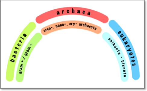 The three life domains
The life phylogenetic tree was constructed from comparative analysis of ribosomal RNA sequences1. Eukaryotic cells include all plant and animal life, and their transition is thought to have risen from the more primitive, prokaryotic cells such as bacteria and archaea through an “endosymbiosis” event.
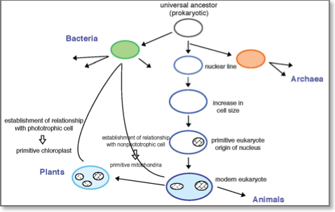
2. Endosymbiotic theory
The hypothesized theory of endosymbiosis deals with the origin of eukaryotic organelles such as the mitochondria in animals and the choloroplasts in plants. According to this theory, certain organelles were originally free-living bacteria that were taken inside another cell as endosymbionts. Mitochondria are one of the many organelles in eukaryotic cells. They are considered to have developed from proteobacteria (in particular, Rickettsiales, the SAR11 clade2, or close relatives). Choloroplasts are the plant counterpart of mitochondria and are considered to have originated from cyanobacteria.
 Summary of the endosymbiotic Theory
Symbiosis occurs when two different species benefit from living together. When one organism lives inside the other it’s called endosymbiosis. It is now generally accepted that mitochondria originated from free-living oxygen-metabolizing (aerobic) bacteria that were engulfed by an ancestral eukaryotic cell that did not make use of oxygen (that is, was anaerobic). Escaping digestion, these bacteria evolved together with the engulfing cell and its progeny, receiving shelter and nourishment in return for the power generation they performed for their hosts, resulting in a permanent relationship. Over years of evolution, mitochondria and chloroplasts have become more specialized and today they cannot live outside the cell.
By now adequate evidence supports this theory, and, in fact, mitochondria and chloroplasts have striking similarities in structure, reproduction and biochemistry. The fact that mitochondria and chloroplasts carry their own genetic information argues in favor of this theory. Each of these organelles has a single, circular DNA that is more similar to prokaryotes than eukaryotes with no histones associated with it. In addition, transfer RNA, ribosomes, and other molecules involved into transcription and translation processes found to be more similar to their prokaryotic counterparts in term of nucleotide sequence, size and even sensitivity to certain antibiotics. Furthermore, the existence of double membranes over many of these organelles suggests the possibility that the inner membrane may have belonged to the original prokaryote while the outer membrane may have formed from food vacuoles as the host cell devoured the prokaryote. The inner membrane of these organelles contains enzymes and transport systems that are similar to the plasma membrane of prokaryotes. Mitochrondria and chloroplasts also replicate by a splitting process similar to binary fission in prokaryotes.
See also: Animation — Endosymbiosis (external link)
3. The cell
The cell is the basic structural and functional unit of all living organisms. The word cell comes from the Latin cellula, meaning “a small room”. With the help of simple microscopy, it quickly became clear that living organisms can be classified on the basis of cell structure into two groups: the eukaryotes and the prokaryotes. The names originate in the Greek word karyon meaning “kernel” or “nucleus”; “eu-” means “well” or “truly.” Multicellular organisms are composed of interplaying configurations of differentiated eukaryotic cells, while prokaryotic cells exist as single-celled, except when they exist in colonies. Eukaryotes keep their DNA in a distinct membrane-enclosed intra- cellular compartment called the nucleus.. Prokaryotes, on the other hand, have no distinct nuclear compartment to house their DNA. Plants, fungi, animals, and the unicellular yeasts are eukaryotes; bacteria and archaea are prokaryotes.
3.1 the prokaryotic cell
Most prokaryotic cells are small and simple in appearance, and they live mostly as independent individuals or in loosely organized communities, rather than as multicellular organisms. They are typically spherical or rod-shaped and measure a few micrometers in linear dimension. Prokaryotic cells often have a tough protective coat, called a cell wall, beneath which a plasma membrane encloses a single cytoplasmic compartment containing DNA, RNA, proteins, water and many other organic and inorganic molecules. The defining characteristic is the absence of a nucleus, however, prokaryotes do possess some internal structures, such as cytoskeletons.Under the electron microscope, this cell interior appears as a matrix of varying texture without any discernible internal organization2. The genomes of prokaryotes are held within an irregular DNA/protein complex in the cytosol, which is a single loop of stable chromosomal DNA stored in an area named the nucleoii that lacks a nuclear envelope. Some bacteria contain a single circular DNA molecule that makes up their entire genome (plasmid), however recent studies have indicated that some prokaryotes contain as many as four linear or circular chromosomes. For example, Vibrio cholerae, the bacterium that causes cholera, contains two circular chromosomes.
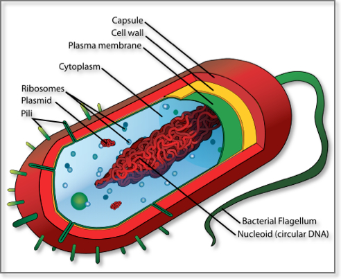 Artistic rendering of a Prokaryotic Cell
3.2 The Eukaryotic Cell
Eukaryotic cells are typically much larger than those of prokaryotes. The major difference between prokaryotes and eukaryotes is that the latter contain various membrane-bound organelles and structures in which specific metabolic activities take place. The most important distinction is the presence of an organelle known as the cell nucleus, a membrane-delineated compartment that houses the eukaryotic cell’s genetic material, the DNA. The eukaryotic DNA is organized in one or more linear molecules, called chromosomes. In humans the nuclear genome contains 23 pairs of chromosomes: 22 pairs of autosomes and one pair of sex chromosomes, giving a total of 46 per cell. Chromosomes are packaged by histones, into a condensed structure called chromatin. Histones serve not only to package the DNA, but also to control its functions3. Chromatin can be in two different forms: a loose form, called euchromatin which consists of DNA that is active and accessible to transcription factors and a more condensed form, called heterochromatin that doesn’t allow transcriptions factors to approach and therefore is inactive4.
In addition to the nucleus, eukaryotic cells include a variety of membrane-bound structures, collectively referred to as the endomembrane system. Simple compartments, called vesicles or vacuoles, can form by budding off other membranes. Many cells ingest food and other materials through a process of endocytosis, where the outer membrane invaginates and then pinches off to form a vesicle. It is likely that most other membrane-bound organelles are ultimately derived from such vesicles. A double membrane (nuclear envelope) with pores that allow material to move in and out surrounds the nucleus. Various tube- and sheet-like extensions of the nuclear membrane form what is called the endoplasmic reticulum (ER), which is involved in protein transport and maturation. It includes the rough ER where ribosomes are attached to synthesize proteins, which enter the interior space or lumen. Subsequently, they generally enter vesicles, which bud off from the smooth ER. In most eukaryotes, these protein-carrying vesicles are released and further modified in stacks of flattened vesicles, called Golgi bodies.
Vesicles may be specialized for various purposes. For instance, lysosomes contain enzymes that break down the contents of food vacuoles, and peroxisomes are used to break down peroxide, which is toxic otherwise. Many protozoa have contractile vacuoles, which collect and expel excess water, and extrusomes, which expel material used to deflect predators or capture prey. In multicellular organisms, hormones are often produced in vesicles. In higher plants, most of a cell’s volume is taken up by a central vacuole, which primarily maintains its osmotic pressure.
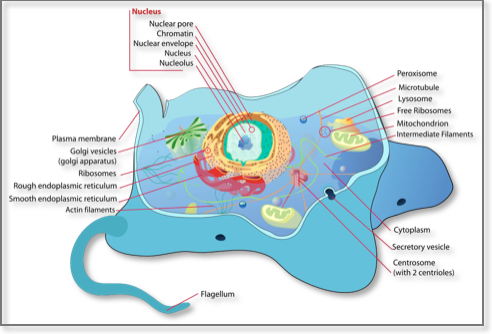 Artistic rendering of a eukaryotic animal cell
3.3 Cellular Processes
Cells have to carry out several functions in order to survive. These functions range from replication to protein synthesis and the generation and recognition of various signals used in cellular communication. Some of these functions are fundamental and their implementation requires complicated cascades of biochemical reactions. Some of the most important processes are described below.
- Reproduction: In order for all living creatures to survive, cells must be able to reproduce. Cells can reproduce in two ways, mitosis and meiosis. Meiosis occurs only in gametes (reproductive cells). During meiosis, the genetic material of the parent cell, the DNA, undergoes reshuffling and results in genetically unique offspring, an important attribute for the maintenance of genetic diversity. Mitosis is used by somatic cells. In mitosis, the resulting daughter cell is identical to the original cell.
- Cellular signaling: The human body consists of approximately 100 trillion (1014) cells. The cell is the basic structural and functional unit of all living organisms. In order for the cells to cooperate, cells need to be able to communicate with each other (intercellular signaling) and to transmit signals into and within the cell (intracellular signaling). Cell signaling is achieved through physical contact (receptor mediating signaling) and through secretion of chemical signals (chemical signaling). From a different point of view, the signal may be mediated via direct contact (juxtacrine signaling), over short distances (paracrine signaling), over larger distances (endocrine signaling) or may target the cell itself (autocrine signaling). There are a large number of intracellular signaling pathways responsible for transmitting information within the cell. They fall into two main categories. The majority responds to external stimuli arriving at the cell surface, usually in the form of a chemical signal (neurotransmitters, hormone, or growth factor), which is received by receptors at the cell membrane, such as the JAK/STAT, the WNT, the hedgehog and the Smad signaling pathways. The other categories are the pathways that are activated by signals generated from within the cell. These are a number of metabolic messengers that act from within the cell to initiate a variety of signaling pathways, such as the Notch signaling pathway. Cellular responses to extracellular stimulation via signal transduction leads to metabolism alterations and gene activation (or deactivation). Gene activation may lead to further cellular effects, if the products of the responding genes include instigators of activation such as transcription factors initiating a cascade of events.
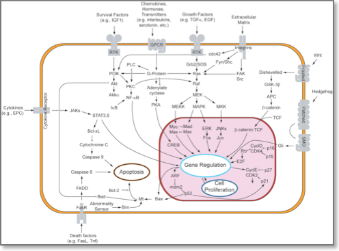 Signal transduction pathways
- Molecular support: For the transport of nutrients and other molecules two main categories exist active transport and passive transport. Small molecules such as oxygen can easily cross the membrane via passive transport in the form of simple diffusion through a concentration gradient. Active transport requires energy. An example of an active transporter is the Na+-K+ ATPase pump. Macromolecules such as proteins, polynucleotides and polysaccharides are also being transferred by active transport.
- Metabolism: All the above-mentioned chemical reactions require energy. The specific details differ in cell types (for example plant cells derive their energy from photosynthesis whereas human cells rely on aerobic respiration) but the end goal is the same i.e. to produce ATP molecules. ATP is a specific nucleotide that is used as a carrier of chemical energy. ATP is adenine with three phosphate groups attached: the bonds connecting the phosphate molecules are highly energetic bonds. When ATP is broken down by hydrolysis, it yields adenosine diphosphate (ADP), inorganic phosphorus and energy. It can also be broken down further into adenosine phosphate (AMP), inorganic phosphorus and energy. The excess phosphorus yielded can be used later for more ATP production from AMP.
4. Non-living genomes of interest
(Adapted from http://en.wikipedia.org/wiki/Virus) A virus is a generally small infective agent that comprises one or more nucleic acid molecules inside a protective protein coat and which can only replicate inside the cells of a living organism. The genetic material is generally either DNA or RNA (never both). In some cases, an envelope of lipids can surround the protein coat when the virus is outside a cell. The shapes of viruses range from simple helical and icosahedral forms to more complex structures. The average virus is about one one-hundredth the size of the average bacterium (approximately 15-25 nanometers in diameter), and, thus, too small to be seen directly with a light microscope. Viral infections in animals provoke an immune response that usually eliminates the infecting virus. Immune responses can also be induced by vaccines, which confer an artificially acquired immunity to the specific viral infection. However, some viruses including those that cause AIDS and viral hepatitis evade these immune responses and result in chronic infections. Antibacterial substances have no effect on viruses, but several antiviral drugs have been developed. The evolutionary origins of viruses are unclear; they may have evolved from plasmids or from bacteria.
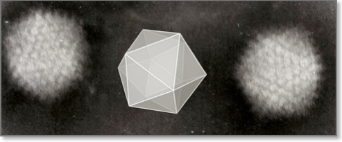 Electron micrograph of icosahedral adenovirus
References
- Jonathan Irving Lunine, C.J.L. Earth: Evolution of a Habitable World, (Cambridge University Press, 1999)
- Bruce Alberts, A.J., Julian Lewis, Martin Raff, Keith Roberts, and Peter Walter. Molecular Biology of the Cell, ( Garland Science, New York, 2002)
- Fischle, W., Wang, Y. & Allis, C.D. Histone and chromatin cross-talk. Current opinion in cell biology 15, 172-183 (2003)
- Grewal, S.I. & Moazed, D. Heterochromatin and epigenetic control of gene expression. Science 301, 798-802 (2003)